by admin | Jul 17, 2015 | Uncategorized

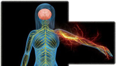
Complex regional pain syndrome (CRPS) is a chronic, predominantly neuropathic and partly musculoskeletal pain disorder often associated with autonomic disturbances. It is divided into 2 types, reflecting the absence or presence of a nerve injury.
Patients with either type may exhibit symptoms such as burning pain, hyperalgesia, and/or allodynia with an element of musculoskeletal pain. CRPS can be distinguished from other types of neuropathic pain by the presence of regional spread as opposed to a pattern more consistent with neuralgia or peripheral neuropathy. Autonomic dysfunction (such as altered sweating, changes in skin color, or changes in skin temperature); trophic changes to the skin, hair, and nails; and altered motor function (such as weakness, muscle atrophy, decreased range of motion, paralysis, tremor, or spasticity) also can be present.1,2
At least 50,000 new cases of CRPS are diagnosed in the United States annually.1 Although the incidence rate is subject to debate, a large epidemiologic study from The Netherlands involving 600,000 patients suggests an incidence of 26.2 per 100,000 individuals. The study also found that women are 3 times more likely to be affected, with postmenopausal women having the greatest risk.3
Presentation
Type 1 CRPS, formerly known as reflex sympathetic dystrophy, often is triggered by a minor or major trauma—fractures account for about 60% of cases.2 Surgery is the next most common precipitating event at 20%. Other etiologies include injections, venipuncture, infections, burns, cerebrovascular accidents, or myocardial infarctions.2,4 There are no identifiable precipitating events in about 10% of patients.2
Type 2, formerly known as causalgia, often is related to high-velocity, blunt injuries, which make up more than 75% of cases. But any process that results in nerve injury, such as surgery, fractures, or injections, also can cause type 2 CRPS.4,5 More than 50% of type 2 cases involving the upper extremities often are related to injuries of the median nerve alone or in combination with another nerve of the upper extremity.5 About 60% of cases in the lower extremities are related to injury of the sciatic nerve.5 Almost all cases involve only partial nerve transection, with upper extremity involvement more prevalent than lower extremity.
Pathophysiology
Historically, CRPS has been poorly understood, and a lack of consistent diagnostic criteria often has been cited in literature. But research in recent years has provided substantial insight into the pathophysiology of the disorder.
As with many other complex conditions, the mechanisms involved in CRPS are multifactorial (Table 1) and include the peripheral and central nervous systems (Figure 1).1 Factors such as altered sympathetic and catecholaminergic function, peripheral and central sensitization, peripheral and central neurogenic inflammation, altered somatosensory representation in the brain, genetics, and psychology all affect patients to varying degrees.
| Table 1. Summary of Pathophysiologic Mechanisms That May Contribute to CRPS1 |
| Altered cutaneous innervation |
- Density of C- and Aδ-fibers in CRPS-affected region
- Altered innervation of hair follicle and sweat glands in CRPS-affected limb
|
| Central sensitization |
- Increased windup in CRPS patients
|
| Peripheral sensitization |
- Local hyperalgesia in CRPS-affected vs unaffected extremity
- Increased mediators of peripheral sensitization
|
| Altered SNS function |
- Bilateral reductions in SNS vasoconstrictive function predict CRPS occurrence prospectively
- Vasoconstriction to cold challenge is absent in acute CRPS but exaggerated in chronic CRPS
- Sympatho-afferent coupling
|
| Circulating catecholamines |
- Lower norepinephrine levels in CRPS-affected vs unaffected limb
- Exaggerated catecholamine responsiveness because of receptor up-regulation related to reduced SNS outflow
|
| Inflammatory factors |
- Increased local, systematic, and cerebrospinal fluid levels of proinflammatory cytokines, including TNF-α, IL-1β, -2, and -6
- Decreased systemic levels of anti-inflammatory cytokines (IL-10)
- Increased systemic levels of proinflammatory neuropeptides, including CGRP, bradykinin, and substance
- Animal postfracture model of CRPS-1 indicates that substance P and TNF-α contribute to key CRPS features
|
| Brain plasticity |
- Reduced representation of the CRPS-affected limb in somatosensory cortex
- These alterations are associated with greater pain intensity and hyperalgesia, impaired tactile discrimination, and perception of sensations outside of the nerve distribution
|
| Genetic factors |
- In the largest CRPS genetic study to date (150 CRPS patients), previously reported associations were confirmed between CRPS and HLA-related alleles
- A TNF-α promoter gene polymorphism is associated with “warm CRPS”
|
| Psychological factors |
- Greater preoperative anxiety prospectively predicts CRPS symptomatology after total knee arthroplasty
- Emotional arousal has a greater effect on pain intensity in CRPS than in non-CRPS chronic pain, possibly via associations with catecholamine release
|
| CGRP, calcitonin gene-related peptide; CRPS, complex regional pain syndrome; IL, interleukin; SNS, sympathetic nervous system; TNF, tumor necrosis factor |
|

Figure 1. Speculative model of interacting CRPS mechanisms.
CGRP, calcitonin gene-related peptide; CRPS, complex regional pain syndrome; IL, interleukin; TNF, tumor necrosis factor
Central Sensitization
Central sensitization is an increased firing of nociceptive fibers in response to intense or persistent noxious stimuli. It is mediated by the nociception-induced release of neuropeptides such as bradykinin, glutamate, and substance P. This phenomenon is part of the reason patients experience allodynia and hyperalgesia. It is not known whether central sensitization precedes, occurs with, or follows development of other CRPS signs and symptoms.1,4,6
Peripheral Sensitization
Peripheral sensitization occurs when an initial tissue trauma causes proinflammatory neuropeptides to be released from primary afferent fibers. These neuropeptides increase background firing of nociceptors, decrease the firing threshold for thermal and mechanical stimuli, and increase firing in response to nociceptive stimuli. This decrease in firing threshold contributes to patients experiencing allodynia and hyperalgesia.1,4,6
Sympathetic Nervous System
Several human studies demonstrated expression of adrenergic receptors on nociceptive fibers after nerve injury. Given that there is likely some sort of nerve damage in type 1 CRPS, this explains why sympathetic outflow has an important effect on pain in patients with this condition. Manipulations with whole-body cooling and heating have supported the theory of sympatho-afferent coupling, although it would not be correct to imply that altered sympathetic function is solely responsible for the development of CRPS. Vascular abnormalities seen in CRPS also are mediated by altered levels of endothelin-1, nitric oxide synthase, nitric oxide, and impaired endothelial-related vasodilatory function. During the progression from acute to chronic CRPS, patients have intense vasoconstriction response in the setting of lowered levels of norepinephrine, implying altered local sympathetic outflow. This is believed to occur due to up-regulation of noradrenergic receptors as a response to low levels of catecholamines. When patients experience pain or regular life stress, these sensitive receptors respond intensely to the release of catecholamines, resulting in a cold, blue, and sweaty appearance.1
Inflammation
The inflammatory process is involved in at least the acute phase of CRPS. There are 2 potential sources of inflammation:
- Classic mechanisms through actions of immune cells, which, after tissue trauma, secrete proinflammatory cytokines, including interleukin-1, -2, and -6, and tumor necrosis factor-α.
- Neurogenic inflammation, which occurs through the release of proinflammatory mediators directly from injured nociceptive fibers in response to various stimuli. These neuropeptides include substance P, calcitonin gene-related peptide, and bradykinin, which promotes plasma extravasation and local tissue edema.1
A subset of patients with CRPS has been found to have low levels of anti-inflammatory and high levels of proinflammatory cytokines.4
Autoimmunity
It has been suggested that autoantibodies may play a role in CRPS. Autoantibodies found in the plasma of patients with CRPS are active at the muscarinic cholinergic and β2 adrenoceptors. Transfer of serum immunoglobulin G to mice from patients with CRPS elicited symptoms of CRPS in recipient mice.7
Genetics
Although there is no clear evidence of genetic predisposition to developing CRPS, it would be prudent to further investigate genetic factors that influence inflammatory and other mechanisms contributing to the syndrome. The largest study of 150 CRPS patients has found a link between CRPS and HLA-related alleles.1,4
Psychological Factors
To date, no evidence has suggested a purely psychological form of CRPS. However, poor coping and emotional stress can certainly raise levels of circulating catecholamines, which could exacerbate vasomotor signs of CRPS, cause pain, and maintain central sensitization.1,4
Brain Plasticity
Functional magnetic resonance imaging scans have shown that there is significant cortical reorganization of somatosensory cortex, which may underlie various manifestations of CRPS. Functional disturbances in posterior parietal cortex responsible for integrating various external stimuli and constructing real-time body schema in space, also may contribute to chronification of pain.1,4,7
Diagnosis
The diagnosis of CRPS is clinical and depends on patient history, physical examination, and findings of musculoskeletal degeneration and secondary pain that develops as a result of persistence of the disease state. CRPS is a diagnosis of exclusion and cannot be made in the presence of other diagnoses that can be responsible for the presentation. Chronic CRPS needs to have symptoms and signs consistent with time-dependent effect of CRPS (ie, atrophy, dystrophy, contractions, and secondary pain).
In the initial several months of CRPS, hypoesthesia and hyperalgesia are common, whereas ongoing disease anesthesia dolorosa can be seen.2 The pain present in later cases when compared with the acute phase is more often present at rest and resistant to treatment. One of the hallmarks of persistent CRPS is the accumulation of orthopedic and neuropathic findings due to altered biomechanics of the affected area and tissue dystrophy and atrophy in superficial and deeper tissues, as well as development of secondary pain in the contralateral limb and other parts of the body as the patient attempts to compensate.
A main reason why CRPS is difficult to diagnose and treat is because the majority of patients do not have classic “warm” (acute) or “cold” (cold) affected limbs—they fall somewhere along the spectrum.1,4 Veldman et al described more patients as having the “cold” type as the duration of CRPS increases, however, some patients with CRPS for more than 10 years still have “warm” limbs.2 And despite being classified as 2 types, there is no evidence that pathophysiologic mechanisms or treatment responsiveness differ in any appreciable way except that type 2 and underlying nerve injury may need to be addressed directly, sometimes surgically (Figure 2).

Figure 2. Schematic representation of treatment modalities based on pathophysiologic mechanisms.7
Skin biopsies of patients with type I CRPS show a significantly decreased amount of C and Aδ-fibers after tissue injury, possibly indicating nerve damage may be present in type 1.1 However, there is no clear evidence as to whether this is a cause or effect of CRPS. Animal models support an increased nociceptive firing in response to norepinephrine, providing evidence that there is sympatho-afferent coupling, which has been suggested by human studies.1 There is further suggestion in animal models that a transcription factor, nuclear factor-β, could play a role in CRPS. This may provide an upstream link between increased proinflammatory neuropeptides and increased proinflammatory cytokines in CRPS.1,4
It also has been shown that patients with CRPS and people with prolonged immobilization, from things like casts for limb fractures, show similar signs of edema, skin color changes, limited range of motion, and altered sensation. This suggests that patients with CRPS experience derangement of normal physiologic responses, making it difficult to identify when these physiologic changes becomes pathologic CRPS and not another diagnosis.4
Diagnostic Tools
During a consensus workshop in 1994, the International Association for Study of Pain (IASP) proposed diagnostic criteria based on clinical symptomatology (Table 2).8 Criticisms of the IASP criteria included a lack of specificity and misdiagnosing other types of neuropathic pain conditions as CRPS. The false diagnosis was thought to stem from the IASP criteria being met solely by self-reported symptoms uncovered by the history without physical signs and symptoms.7
| Table 2. IASP CRPS Diagnostic Criteria8 |
2-4 of the following with 2, 3, and 4 being mandatory:
- The presence of an initiating noxious event, or a cause of immobilization.
- Continuing pain, allodynia, or hyperalgesia with which the pain is disproportionate to any inciting event.
- Evidence at some time of edema, changes in skin blood flow, or abnormal sudomotor activity in the region of the pain.
- This diagnosis is excluded by the existence of conditions that would otherwise account for the degree of pain and dysfunction.
|
All of the following:
- The presence of continuing pain, allodynia, or hyperalgesia after a nerve injury, not necessarily limited to the distribution of the injured nerve.
- Evidence at some time of edema, changes in skin blood flow, or abnormal sudomotor activity in the region of the pain.
- This diagnosis is excluded by the existence of conditions that would otherwise account for the degree of pain and dysfunction.
|
| CRPS, complex regional pain syndrome; IASP, International Association for the Study of Pain |
|
In an aim to improve the IASP criteria, an international consensus meeting was held in Budapest in 2003. The results were based on the previously published Harden/Bruehl criteria (Table 3).9 A 2010 study showed the IASP criteria of being 100% sensitive but only 41% specific in 113 CRPS type 1 patients and 47 non-CRPS neuropathic pain patients. The new Budapest criteria revealed 99% sensitivity with 68% specificity.10 Veldman’s criteria includes physical signs in combination with symptoms and was derived from a cross-sectional cohort study of 829 patients (Table 4).2
| Table 3. Harden/Bruehl CRPS Diagnostic Criteria9 |
- Continuing pain, which is disproportionate to any inciting event
- Must report ≤1 symptom in 3 of the following 4 categories:
- Sensory:
Reports of hyperesthesia and/or allodynia
- Vasomotor:
Reports of temperature asymmetry and/or skin color changes and/or skin color asymmetry
- Sudomotor/Edema:
Reports of edema and/or sweating changes and/or sweating asymmetry
- Motor/Trophic:
Reports of decreased range of motion and/or motor dysfunction (weakness, tremor, dystonia) and/or trophic changes (hair, nails, skin)
- Must display ≤1 sign at time of evaluation in ≥2 of the following categories:
- Sensory:
Evidence of hyperalgesia (to pinprick) and/or allodynia (to light touch and/or temperature sensation and/or deep somatic pressure and/or joint movement)
- Vasomotor:
Evidence of temperature asymmetry (>1∞C) and/or skin color changes and/or asymmetry
- Sudomotor/Edema:
Evidence of edema and/or sweating changes and/or sweating asymmetry
- Motor/Trophic:
Evidence of decreased range of motion and/or motor dysfunction (weakness, tremor, dystonia) and/or trophic changes (hair, nails, skin)
- There is no other diagnosis that better explains the signs and symptoms
|
| Same as CRPS I but with the evidence of a peripheral or central nerve injury |
| Patients who do not fully meet the clinical criteria, but whose signs and symptoms cannot be better explained by another diagnosis |
| CRPS, complex regional pain syndrome; NOS, not otherwise specified |
|
| Table 4. Veldman2 CRPS Diagnostic Criteria |
- Presence of 4 or 5 of the following:
- Unexplained diffuse pain
- Difference in skin color relative to other limb
- Diffuse edema
- Difference in skin temperature relative to other limb
- Limited active range of motion
- Occurrence or increase of above signs and symptoms after use.
- Above signs and symptoms are present in an area larger than the area of primary injury or operation and include the area distal to the primary injury.
|
|
| CRPS, complex regional pain syndrome |
|
Treatment
Although the majority of CRPS symptoms resolve within an approximate 12-month period, an estimated 25% of patients still fulfill IASP diagnostic criteria at 12 months and may suffer from CRPS chronicity. CRPS following fracture has a better resolution rate; “cold” CRPS or upper-limb involvement has the worst outcome. Because CRPS is a multifactorial disease with poorly understood mechanisms, the mainstay of treatment remains physical and occupational therapy aimed at return and preservation of function, prevention of loss of range of motion, and prevention of contractures and atrophy.4
Pharmacologic Treatment
IV Ketamine has proven to be very effective in the treatment of CRPS /RSD
At the Florida Spine Institute, treatment protocols are individually planned depending on the nature of pain and the patient’s responsiveness to initial sessions. Infusion cocktails are prepared in house so that they can be tailored to each patient’s therapeutic needs. A variety of medications are often used:
- Lidocaine
- Ketamine
- Bisphophonates
- Magenesium
These medications are typically mixed with saline in an IV bag and infused slowly over several hours, depending on the medication and/or protocol being used. Usually, a series of treatments will be recommended daily for a period of a week or more. The duration of pain relief following one or more ketamine infusions cannot be predicted. The goal is to achieve lasting relief as measured in weeks or months following the last treatment. Most patients who enjoy prolonged pain relief will need to return on occasion for a booster infusion, or continue to take low dose intranasal ketamine at home.
There is also convincing results with regard to IV bisphosphonates—most recently neridronate in patients with disease duration of less than 6 months. At 1-year follow-up, neridronate showed improved pain control. Multiple neuropathic medications such as gabapentin, tricyclic antidepressants, and opioids have been used through their extrapolated benefit in neuropathic conditions other than CRPS. Oral steroids continue to be used in acute CRPS, although the evidence is poor and sympatholytic drugs are used by clinicians with low success rates.7,11,12 But a recent small trial using low-dose oral phenoxybenzamine showed significant functional improvement in patients with CRPS.13 Taking everything above into consideration, we can conclude that the majority of pharmacologic treatments used by clinicians are quite empirical and largely based on personal preferences and experiences.
Interventional and Surgical Techniques
The main utility of interventional pain medicine in CRPS is to enable proper physical or occupational therapy and break the cycle of peripheral and/or central pain. Moderate evidence shows that sympathetic blockade is effective. Spinal cord stimulators appear to provide significant improvement of function in type 1 CRPS, and are more cost-effective over a patient’s lifetime compared with physical therapy and medical management.4,14 Although there is great resistance to surgery for CRPS patients, significant pain resolution may be achieved through nerve decompression or denervation procedures, neuroma resection, and neurolysis once patients are properly identified by nerve blocks. In many patients, the noxious stimulus is maintained through nerve compression; osteophytes; fibrosis; neuroma; arteriovenous malformation; or anything that entraps, compresses, or distorts the nerve. Surgery may be indicated in these cases.
Psychological Interventions
Psychological factors play a role in the treatment of CRPS. There is likely benefit in cognitive-behavioral therapy. Correcting body image also may help in CRPS affected patients.4,7
References
- Bruehl S. An update on the pathophysiology of complex regional pain syndrome. Anesthesiology. 2010;113(3):713-725.
- Veldman PH, Reynen HM, Arntz IE, et al. Signs and symptoms of reflex sympathetic dystrophy: prospective study of 829 patients. Lancet. 1993;342(8878):1012-1016.
- de Mos M, de Bruijn AGJ, Huygen FJPM, et al. The incidence of complex regional pain syndrome: a population-based study. Pain. 2007;129(1-2):12–20.
- Borchers AT, Gershwin ME. Complex regional pain syndrome: a comprehensive and critical review. Autoimmun Rev. 2014;13(3):242-265.
- Hassantash SA, Afrakhteh M, Maier RV. Causalgia: a meta-analysis of the literature. Arch Surg. 2003;138(11):1226-1231.
- Rockett, M. Diagnosis, mechanisms and treatment of complex regional pain syndrome. Curr Opin Anaesthesiol. 2014; 27(5):494-500.
- Gierthmühlen J, Binder A, Baron R. Mechanism-based treatment in complex regional pain syndromes. Nat Rev Neurol. 2014;10(9):518-528.
- Merskey M, Bogduk N, eds. Classification of chronic pain: descriptions of chronic pain syndromes and definition of pain terms; second edition. Seattle: WA; 1994.
- Harden RN, Bruehl S, Stanton-Hicks M, et al. Proposed new diagnostic criteria for complex regional pain syndrome. Pain Med. 2007;8(4):326-331.
- Harden RN, Bruehl S, Perez RS, et al. Validation of proposed diagnostic criteria (the “Budapest Criteria”) for complex regional pain syndrome. Pain. 2010; 150(2):268-274.
- Rowbotham, MC. Pharmacologic management of complex regional pain syndrome. Clin J Pain. 2006:22(5):425-429.
- O’Connell NE, Wand BM, McAuley J, et al. Interventions for treating pain and disability in adults with complex regional pain syndrome. Cochrane Database Syst Rev. 2013;4:CD009416.
- Inchiosa MA Jr. Phenoxybenzamine in complex regional pain syndrome: potential role and novel mechanisms. Anesthesiol Res Pract. 2013;2013:978615.
- Taylor RS, Van Buyten JP, Buchser E. Spinal cord stimulation for complex regional pain syndrome: a systematic review of the clinical and cost-effectiveness literature and assessment of prognostic factors. Eur J Pain. 2006;10(2):91-101.

by admin | Jul 17, 2015 | Uncategorized
Researchers Continue To Look for Basic Understanding of CRPS Causes

Researchers tasked with developing a rudimentary understanding of complex regional pain syndrome (CRPS) are dividing their attention in several different directions. According to several experts who spoke at the 2013 International Congress on Neuropathic Pain, there is evidence for inflammatory, neuropathic and immunologic roots to the enigmatic syndrome, and further investigation into these three aspects of the syndrome is necessary for the development of more effective treatments.
“These different contributing factors all influence each other, so we need to address all of them so that patients don’t get onto a downward spiral where each factor worsens another,” Anne Louise Oaklander, MD, PhD, associate neurologist at Massachusetts General Hospital and associate professor at Harvard Medical School, both in Boston, told Pain Medicine News after attending the panel discussion.
Not a Perfect Fit
Ralf Baron, MD, vice chair of the Department of Neurology and head of the Division of Neurological Pain Research and Therapy at the University Hospital Schleswig-Holstein in Kiel, Germany, told attendees that until recently, CRPS was understood to be clearly a neuropathic pain disorder. However, CRPS does not fit with the 2008 redefinition of neuropathic pain, defined as “pain arising as a direct consequence of a lesion or disease affecting the somatosensory system” (Neurology 2008;70:1630-1635).
“CRPS is neuropathic in that there are characteristic neuropathic sensory abnormalities, but it also shows signs of central sensitization, inflammation, and autonomic and motor abnormalities,” Dr. Baron told attendees.
One way of grouping CRPS patients is by looking at their distinct somatosensory dysfunctions, Dr. Baron suggested. Individuals with deficits in temperature detection but no allodynia, and with loss of small nerve fibers, innervation and nerve degeneration, can be classified as having a neuropathic disorder. A second cluster of patients can be seen as having central sensitization, with normal temperature sensitivity but severe mechanical and thermal hyperalgesia. A third patient cluster may have inflammatory CRPS, with deep hyperalgesia and heat hyperalgesia but no hyperalgesia to prick testing, Dr. Baron explained.
An Autoimmune Disease?
Another way of understanding CRPS, proposed by Andreas Goebel, MD, PhD, senior lecturer and honorary consultant at the University of Liverpool and Walton Centre National Health Service Foundation Trust in Liverpool, United Kingdom, is that a subset of CRPS patients have an autoimmune disorder–related condition.
“It is possible that a regional immune response is triggered following stress, inflammation and trauma,” he posited.
Dr. Goebel believes some of the 15% of CRPS patients with refractory symptoms lasting longer than six to 12 months may fall into this group (Pain 2009;142:218-224). He noted that several studies support the autoimmune paradigm, with results showing that a subset of CRPS patients have elevated levels of serum antibodies to several bacterial pathogens.
“Furthermore, there is evidence for CRPS serum immunoglobulin binding to peripheral nerves,” he said (Neurology 2004;9:1734-1736).
Indeed, several small case series, including his own, have demonstrated the efficacy of intravenous immunoglobulin G (IgG) in subsets of patients with long-standing, refractory CRPS, Dr. Goebel said (Pain Med 2002;3:119-127). Additionally, a randomized controlled trial of 12 patients with long-standing CRPS who received the agent found that 25% had pain relief at least 50% relative to baseline and 17% had improvements in pain between 30% and 50% (Ann Intern Med 2010;152:152-158).
Although IgG has anti-inflammatory effects in addition to being an immunomodulator, Dr. Goebel believes IgG’s efficacy is likely not explained by its anti-inflammatory effect.
“All of the investigations which we have done, both in the lab and clinically, have been leaning more and more toward confirming there is an autoimmune aspect to CRPS,” Dr. Goebel concluded. “However, since we do not know which structures the autoimmune response is targeting, our current evidence remains somewhat indirect.”
According to Dr. Goebel, if some patients in fact have CRPS of autoimmune origin, “a range of potential therapies, such as therapeutic plasma exchange and B-cell modulating therapies, can be at our disposal.
“These have all been tried and tested in other autoimmune disorders, and we can have access to an armamentarium that we did not have before,” he said.
An “Ultralocal” Inflammatory Response?
Frank Huygen, MD, PhD, professor of anesthesiology at Erasmus Medical Center in Rotterdam, the Netherlands, argued the inflammatory component to CRPS could be the most clinically meaningful element of the syndrome in some patients. However, rather than looking for systemic inflammation, he believes researchers need to consider an “ultra-local” inflammatory response.
“There are inflammatory mediators that are increased in the blister fluid of an involved extremity in CRPS,” Dr. Huygen said.
His own research has documented increased levels of interleukin-6 and tumor necrosis factor (TNF)-alpha in some patients (Eur J Pain 2008;12:716-721).
In a small, placebo-controlled, randomized trial, Dr. Huygen and his colleagues showed that the anti-TNF drug infliximab (Remicade, Janssen) exacerbated symptoms in some CRPS patients, leading to discontinuation of the trial (Pain Pract 2013; May 22: doi: 10.1111/papr.12078). The researcher still believes there could be a role for infliximab and other biologics in the treatment of CRPS.
“Although the sponsor stopped the study early, preliminary data showed enormous reductions in TNF-alpha levels in blister fluid with infliximab treatment, but these did not correlate with clinical changes,” he said.
Summarizing the challenge of understanding and treating a complex syndrome, Dr. Baron suggested CRPS should no longer be seen monolithically as a neuropathic disorder.
“As long as we do not have a clearer pathophysiological picture of CRPS patients, and because of the obvious heterogeneity of the signs, symptoms and mechanisms of the syndrome,” he said, “it would be wise to look at the condition separately from other classical neuropathic pain syndromes.”
—David Wild
by admin | Jul 17, 2015 | Uncategorized
Relief for Worst RSD May Lie With Ketamine Coma

For the most severe cases of reflex sympathetic dystrophy (RSD), inducing a five-day coma may be the only effective treatment.
The method is akin to rebooting the central nervous systems of patients whose nerve cells have gone haywire. The FDA has yet to approve coma therapy, which is induced by administering large bolus injections of ketamine and midazolam at up to 50 times the normal dose. But that has not stopped two U.S. doctors from pioneering the use of a “ketamine coma” in American patients treated at hospitals in Germany and Mexico.
For the better part of four years, Robert Schwartzman, MD, chairman of the Department of Neurology at Drexel University College of Medicine in Philadelphia, and Anthony Kirkpatrick, MD, PhD, an anesthesiologist at the University of South Florida College of Medicine in Tampa, have been studying the effects of ketamine treatment, including induced comas, in patients with RSD. Their results suggest that the coma therapy may provide long-term and perhaps permanent relief in as many as half of the most severe cases.
Ketamine first won FDA approval in 1970 as an anesthetic. It quickly became known on the street as “Special K,” a powerful hallucinogen similar to LSD and PCP. Clinicians in the United States can legally give the drug to patients with RSD—also known as complex regional pain syndrome (CRPS) type 1—who are under conscious sedation. In this group, relief typically lasts no more than six months, Dr. Schwartzman said.
Potent Agent
Ketamine is the most potent clinically available inhibitor of N-methyl-d-aspartate (NMDA) receptors. These receptors permit the transfer of electrical signals between neurons in the brain and the spinal column. Studies support the idea that RSD results from a dynamic change in the physiology and structure of central pain neurons mediated by NMDA receptors. When these receptors malfunction, enzymatic and metabolic cascades occur in pain cells, and the degree of pain is magnified out of proportion to the physical injury causing it.
In a study of infusions of low-dose ketamine (Pain Physician 2005;8:175-179), Dr. Schwartzman and colleagues found that a critical factor in central sensitization seems to be the release of magnesium on the NMDA receptor, with an influx of calcium and the initiation of intracellular cascades. As an NMDA antagonist, ketamine blocks central sensitization. Drugs such as dextromethorphan, amantadine and memantine (Namenda, Forest Pharmaceuticals) also could be used, but they appear to have a low potential for blocking the sensitization process.
Ketamine has long been known to be able to prevent RSD/CRPS following surgery, said Scott Reuben, MD, professor of anesthesiology and pain medicine at Tufts University School of Medicine in Boston and director of pain management at Baystate Medical Center in Springfield, Mass.
“If it [ketamine] can prevent CRPS, the thought was, ‘can we use it to treat it?’ ” said Dr. Reuben, who serves as an adviser to the Mexico study. “This is just the stepping stone. Unfortunately, all we have are case reports and retrospective studies. We need prospective studies.”
Only the Worst Patients
RSD affects between 200,000 and 1.2 million Americans, according to the Reflex Sympathetic Dystrophy Association. The disorder develops without any apparent explanation in 1% to 2% of patients with fractures and in 2% to 5% of patients with peripheral nerve injuries. The RSD group claims that roughly 50,000 new cases develop each year.
For the Mexican and German research, doctors chose patients with the most severe cases of RSD who had tried everything—including blocks and hyperbaric chambers—for their pain.
Of the roughly 200 to 300 new patients with RSD whom Dr. Kirkpatrick sees each year, fewer than 5% are so unresponsive to conventional therapies that they are considered for treatment with a ketamine coma. Those who are eligible can barely endure even the most innocuous sensations, Dr. Kirkpatrick said. Patients report feeling pain at the slightest touch, from a dog’s wagging tail to air flowing over the skin. One patient lived in a box because it hurt her to wear clothes. “Some of these patients,” Dr. Kirkpatrick said, “are about ready to die.”
Frustrated physicians from around the world refer patients to Drs. Kirkpatrick and Schwartzman. “I only see the worst patients,” said Dr. Schwartzman, who took on the challenge of RSD after being unable to cure one of his patients with the condition. “Some have gone through up to 100 doctors.”
Failure in More Than Half
Dr. Schwartzman has sent a total of 38 patients to Germany for treatment with the ketamine coma, which was discovered serendipitously by Ralph-Thomas Kiefer, MD, and Peter Rohr, MD, in Tübingen. The two physicians had induced a coma in a patient with RSD and severe head trauma. When the patient awoke, the pain syndrome had vanished.
The five-day coma is induced with large bolus injections of ketamine (1-1.5 mg/kg) and midazolam (2.5-7.5 mg). The coma is maintained with infusions of ketamine (3-7 mg/kg per hour) and midazolam (0.15-0.3 mg/kg per hour), which are tapered toward the end.
Every patient in whom a coma is induced does well initially, Dr. Schwartzman said, but the pain returns in 55% to 60% of cases. Still, of the 38 patients treated in Germany, at least 12 have had minimal or no pain for more than five years. Three of the 12 required occasional subanesthetic boosters of ketamine.
“We’re blocking the RSD,” Dr. Schwartzman said. “The maintaining thing is still there. If you don’t block the maintaining problem, the same molecular genetic cascade occurs.”
A study of the full ketamine coma in the patients treated in Germany will soon appear in Pain Medicine. Of the 20 patients featured in the study who underwent the therapy, 16 experienced complete remission lasting at least six months. “While the trial suggests improvements in pain reductions,” the researchers concluded, “a randomized controlled study will be necessary to prove its efficacy.”
The coma’s side effects—precipitous weight loss, sleep disruption, anxiety, weakness and the usual complications of critical care medicine—are potentially serious. The bottom line, Dr. Schwartzman said, is that “the procedure has proven to be very safe but clearly has inherent risks.”
South of the Border
Dr. Kirkpatrick has embarked on a study in Mexico with a protocol similar to that used in the German study; he sends his patients to the San José Technological Hospital, affiliated with the Tec de Monterrey School of Medicine in Monterrey, Mexico, a few hours’ drive from the Texas border. Patients pay about $20,000 for the treatment, which is not covered by insurance.
Leading the Mexican medical team is Fernando Cantœ Flores, MD, an anesthesiologist and specialist in pain management who was trained at the University of Texas.
The study was originally approved in the United States by the institutional review boards of the University of South Florida and Tampa General Hospital, but the FDA refused to grant an exemption to its international new drug application. Rather than embark on a process that would likely cost $3 million and delay treatment for their patients, Dr. Kirkpatrick and his colleagues moved the study to Mexico. A review board in Monterrey also approved the study.
So far, eight patients have been treated in Mexico. The main difference from the German study is that pain thresholds are measured with a force gauge. The German study relied on self-reporting.
Low-Dose Conscious Sedation
A less dramatic treatment option for severe cases of RSD is a subanesthetic infusion of low-dose ketamine (10-30 mg per hour titrated according to side effects such as amnesia, blurred vision and vomiting). Dr. Schwartzman has performed close to 200 of these—about one each week for the past four years—at Hahnemann University Hospital in Philadelphia. The treatment costs about $10,000, and insurance companies will not pay for it. Dr. Schwartzman estimated that he performs 95% of all such procedures in the country. He also treats about 10 outpatients per week with lower doses in his clinic.
The infusions succeed in 70% to 80% of cases. But even in the most responsive patients, pain typically returns after approximately six months. A study published in Pain Medicine (2004;5:263-275) found similar results.
A two-hour infusion of low-dose ketamine can also be used to manage RSD or as a booster after treatment with the coma. Pretreatment with 0.2 mg of I.V. glycopyrrolate (the only other drug needed) is administered along with a ketamine drip at 100 mg per hour, supplemented with 5- to 40-mg I.V. bolus injections of ketamine. An average adult will require a total of 400 to 600 mg of ketamine over a two-hour period.
For patients with the most intractable cases of RSD, the full coma treatment may still be the only hope. In a study published online in Pain Medicine in July 2007, (online early article), Dr. Schwartzman and his German colleagues found that an “awake” version of the ketamine infusion at higher doses (50-500Êmg per day) over a 10-day period in four patients with extreme RSD did not relieve pain.
Cause for Optimism
Shannon Stocker, MD, an Orlando, Fla., RSD patient, underwent coma therapy in Mexico. Dr. Stocker said the concern she felt about going into the treatment was “overwhelmed by a desire to get better because the pain was so bad it was worth all the risk. The burning pain was constant. But when there was anything like raindrops, it felt like ice picks stabbing me.”
The ketamine coma may be the key to curing other conditions directly related to either RSD or similar nerve dysfunction. As a result of her RSD, Dr. Stocker had skin ulcers all over her arms, which began to clear up while she was still in the coma.
Jim Broatch, executive director of the RSD Syndrome Association, called ketamine therapy “promising” but added that more data are needed. “We’re saying the jury is still out.”
Dr. Kirkpatrick agreed. “We’re just trying to do hard science,” he said. “We can’t make progress in this research if we ignore the bad things.”
In January 2008, Dr. Kirkpatrick plans to open The RSD/CRPS Treatment Center and Research Institute in Tampa. An entire city block has already been purchased by the International Research Foundation for RSD/CRPS, which Dr. Kirkpatrick cofounded with a patient.
Dr. Schwartzman has now written more than 60 articles on RSD and spoken about the disorder at more than 100 conferences, including the 2007 annual meeting of the American Academy of Pain Management, at which he delivered the keynote address.
The success of the various ketamine protocols has made him optimistic about the prospects for patients with previously intractable RSD. “It’s wonderful to be able to successfully treat someone in severe pain,” he said. “That’s why you go to medical school.”
by David Rosenfeld