
Introduction
Radiofrequency ablation is a term used when radio waves are used to produce heat to destroy tissue, usually a nerve. It has been used for several years with great success in patients who have abnormally fast heartbeats. More recently, it is being used to destroy tumors. It is a non-surgical option to treat your spine pain.
Spine pain is the second most frequent pain complaint. It occurs in 65 to 80 percent of the population at one time or another, and can be disabling and frightening. Its costs to society are great.
In the case of spinal pain, radiofrequency waves are transmitted through a needle placed into the facet joint under x-ray guidance. This procedure is also known as rhizotomy.
This guide will help you understand
- What parts of the spine are involved
- What is the surgeon is trying to achieve
- What happens during the procedure
- What are the possible complications
Anatomy
What parts make up the spine?
The spine is made up of three general parts. The top portion is the cervical spine and connects with the skull or cranium. The middle portion is the thoracic spine and is identified by the ribs that attach to each of the vertebrae. The lower portion is the lumbar spine. It connects with the pelvis at the sacrum.
The human spine is made up of 24 spinal bones called vertebrae. Vertebrae are stacked on top of one another to form the spinal column. The spinal column is the body’s main upright support.
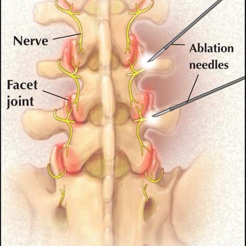
The vertebrae have discs that serve as cushions in between them. Each vertebra has two sets of bony knobs that meet between each vertebra. These form facet joints. The facet joints are located on the back of the spinal column in the lumbar and thoracic spine. In the neck, or cervical spine, they are located on each side of the vertebra. They are also called zygoaphophyseal or apophyseal joints.
A joint is where two or more bones are joined, allowing motion. Facet joints allow flexion or forward bending, and extension or backward bending, as well as rotation of your spine. The facet joints are synovial joints. This means that a capsule of soft tissue encloses them to help support it. It also makes fluid that lubricates the joint, like oil for the moving parts of a machine.
The surfaces of the joints are lined with cartilage. This allows joints to move or glide smoothly. The articular cartilage surface of the facet joint can become thin due to wear and tear. Bone spurs and enlargement of the joint can occur due to chronic inflammation and arthritis.
Nerves called medial branches supply facet joints. They carry the pain signals to the spinal cord. The signals eventually reach the brain where the pain is registered. Pain is a warning when the joint is irritated.
Rationale
What do surgeons hope to achieve with this procedure?
There are several structures in the spine that can be a source of pain. One of the most common sources is the facet joint. The joints can develop arthritis and cause acute and chronic pain. The pain may come and go depending on activity.
Facet joint pain as the cause of back pain can be determined by a facet joint injection. This is called a diagnostic injection, meaning the doctor uses it to help make a diagnosis. The physician using x-ray guidance will inject the facet joint(s) in question with a small amount of a combination of local anesthetic and cortisone. Relief of the acute or chronic problem while the joint is numb indicates that you will likely have a good response to radiofrequency ablation.
Radiofrequency ablation uses radio waves to produce heat to destroy the nerve(s) carrying the pain signals from the facet joint. Once the nerves carrying pain sensation from the joints are destroyed, your pain should be reduced. This should allow you to do more activity, and decrease your pain medications.
Preparation
How should I prepare for the surgery?
Your surgeon will discuss the preoperative guidelines. Follow your surgeon’s instructions. These instructions may include
- Do not eat or drink for at least six hours before the procedure. You will be able to take your usual medication with a small amount of water. If you have diabetes, do not take your insulin or diabetic pills until after the procedure
- You will need a driver to return home
- Do not take any aspirin or aspirin-containing medication at least eleven days before the procedure. They may prolong bleeding
- Wear loose fitting clothing that is easy to take off and put on
- Take a shower the morning of the procedure, using a bactericidal soap to reduce chances of infection
- Do not wear jewelry
Procedure
What happens during the procedure?
When the procedure is to begin, an IV will be started. This will allow the use of medications to help sedate you and make the procedure more comfortable. It is also important to have IV access for medications if you should have an allergic reaction during the procedure.
You should be awake for the procedure to help the doctor with correct placement of the electrode used for radiofrequency ablation. You will not be given a general anesthetic. The area to be treated will be cleaned and then numbed with a local anesthetic.
Using x-ray guidance, the doctor will place the needle in the proper facet joint. A microelectrode is then placed inside the needle. A small radiofrequency current is then sent to the medial branch nerve of the joint capsule for approximately 60 to 90 seconds. The procedure is done with sterile technique to minimize the risk of infection.
After the procedure, you will be taken to a recovery area. The nurses will monitor you and be sure you do not have an allergic reaction. You will be allowed to leave once you are stable.
Complications
What might go wrong?
This procedure is a safe, non-surgical treatment, and the risks for complications are low. However there are several complications that may occur during or after this procedure. No procedure is 100 percent foolproof. This article doesn’t provide a complete list of all the possible complication, but it does highlight some of the most common problems. Complications are uncommon, but you should know what to watch for if they occur. Some of the most common complications following radiofrequency ablation include
- Neuritis
- Neuroma
- Numbness-nerve damage
- Infection
- Allergic reaction
- Lack of pain relief
Neuritis
Neuritis is an inflammation of the nerve with pain and tenderness that lasts three to six weeks. This can occur in 10 to 15 percent of patients. Neuritis usually goes away by itself. If it doesn’t, the doctor can inject a local anesthetic along with a steroid into the nerve. Pulsed radiofrequency can also be used to help with the pain and inflammation.
Neuroma
A neuroma is a tumor from a nerve made of nerve cells and fibers. It forms around the area destroyed during radiofrequency ablation. The tumor can put pressure on the nerve and nearby tissue causing increased pain.
Numbness
Numbness caused by nerve damage can occur. Permanent numbness is also a possible complication.
Infection
Infections can also happen. Infections can involve the skin, or the joint and surrounding bone.
Allergic Reaction
Allergic reactions can occur since medications and sometimes contrast dye is used during the procedure. Anaphylaxis is an allergic reaction that is serious and can result in death.
Lack of Pain Relief
Even though a test block was beneficial, some patients have no pain relief from the procedure.
After Care
What should I expect after surgery?
Immediately following the procedure, you may have some relief of pain from the numbing medication used during the procedure.
You will not be able to drive or do any physical activity for 24 hours.
You may experience an increase in pain for the first several days following the procedure. Additional pain medications may be necessary to make you comfortable. If these include narcotics, you will need to watch for constipation. Drink lots of fluids and eat foods with plenty of fiber. If constipation should occur you will need to use a laxative, available over-the-counter.
You may also note some swelling and bruising where the needle was inserted. Using a cold pack may ease the discomfort. Occasionally infection or bleeding can occur at the site of the procedure. If you have a fever of 101 degrees or greater, chills, or redness or drainage at the treatment site, call your doctor.
The degree of pain relief varies from person to person. The maximum decrease in pain may take up to three or more weeks to occur. You can eventually expect 50 percent or greater pain relief. Pain relief can last from six to12 months or even longer. The nerves do repair themselves and your pain may return. The procedure may be done again.
Your doctor will arrange a follow-up appointment, or phone consult within three to four weeks after the procedure to see how you are doing.
Rehabilitation
What should I expect during my rehabilitation?
It is important that you start a program of conditioning, strengthening, and range of motion exercises after radiofrequency ablation. Ideally, increased muscle strength around the arthritic joints will make them less painful for several months after radiofrequency ablation.